X rays Physics and Roentgens

X rays Physics and Roentgens
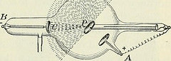
In 1895, Wilhelm Roentgen, a German physicist, was experimenting with an electrically charged cathode tube, called a Crooke’s tube. He wrapped the tube in cardboard to shield out the visible light which it produced. A special chemical plate glowed several feet away. He speculated that the tube had emitted unknown rays which penetrated the cardboard and made it to the plate. He named these unknown rays “x-rays”. In his further experiments he used his wife’s hand as a target, and made a picture of her hand and finger bones on a photographic plate. This was the first x-ray in medical history. It was published in a journal not too long afterward, and the scientific community received it with enthusiasm. Roentgen received the Nobel Prize in Physics in 1903 for his work.
Numerous scientists began to work on variations of the Crooke’s tube which made it more durable and efficient in emitting x-rays (also called Roentgen rays, especially in Germany).
It was found that x-rays could not be diverted with magnetism, but they could be refracted with crystals. This furthered the conclusions that x-rays were energy waves like visible light, only much smaller in width and more powerful. It was found that there were hard x-rays which penetrated deeper and soft x-rays which tended to be absorbed. Like visible light xray travel in particle-like packets called photons. A bone is made largely of calcium which blocks the x-ray; consequently it appears as a white, non-exposed area on the x-ray film. Soft tissues which allow partial penetration show up as grey on the film. The x-rays totally penetrate air structures and appear as black on the film.
Nikolai Tessla was one of the first scientists to warn of the potential adverse effects of x-rays on living tissue. Up until that time a lot of scientists had ended up with cancers of their hands and arms, sometimes requiring amputation. We are now quite careful about keeping track of absorbed dosage which a patient and his x-ray technician receive. The FDA has warned about the overuse of such imaging as the CT scan, which can deliver the x-ray energy of four-hundred single chest x-rays. When I was a child, many major shoe stores had foot fluoroscopes where you could x-ray your own feet to see if your shoes fit. Fortunately these machines were removed from use. The first medical x-rays after the improvements to the x-ray tube were fluoroscopies. These allowed the doctor to look at bones and adjacent structures in motion. Later came the use of photographic silver plates to make still pictures. As mentioned, these plates have largely been replaced by digital receiving apparatus which place and store the images on a computer.
It has only been about thirty years that the CT x-ray device has been patented and first used. The CT utilizes an x-ray tube moving around the patient taking millimeter slice images. These are converted to three-dimensional images by a computer. Again the x-ray dosage of these machines has been the object of recent concern because of the amount of radiation delivered. Several variations of x-ray procedures have been developed to look at soft tissue structures in detail such as arteries, veins, the inner surfaces of air-inclusive organs, and the central nervous system. These include giving contrast media either intravenously, orally, or rectally. This causes the soft tissue structures of interest to stand out as white the way bone normally does. Examples of contrast x-rays would be heart catheterization, contrast head CT, contrast x-ray or CT of the kidneys, or the upper or lower gastrointestinal (G.I.) series.
The MRI, a non x-ray device, has taken precedence over x-rays in many cases because of the absence of radiation. The MRI uses very large magnets to align electrons in tissues to make computer delineated images. These are especially good of the brain and spinal cord, the joints, and in certain other areas where a plain x-ray does not yield adequate information. The drawback is that it is comparatively very expensive and requires a very high technology to accomplish.
If you have a broken bone the x-ray is the procedure of choice. It not only shows such details as angle and displacement of the fracture, but also shows if the fracture is properly fixed with treatment. A plain x-ray of the chest is adequate to diagnose pneumonias and cancers in the lung tissue. The mammogram is very sensitive in looking for abnormal growths in the breasts. Heart catheterization has revolutionized the diagnosis and treatment of heart disease. Sometimes scopes are chosen over certain x-rays, as in colonoscopy being chosen over a lower G.I. x-ray, but there is a place for both procedures. CT of the head is a rapid way to assess brain injury or stroke.
The important consideration to make is whether the benefit of the x-ray outweighs the risk. It is okay for you to ask your physician this question when he suggests or orders an x-ray. In some cases the answer is yes; in others it is no. The judicious use of x-rays is a cornerstone of modern medicine.
John Drew Laurusonis, M.D.
Doctors Medical Center
Dr. Laurusonis was conferred his Doctor of Medicine degree in 1983 and has been actively taking care of patients since completing his Internal Medicine residency in 1987 in the Garden State of New Jersey. Dr. Laurusonis has been licensed in four states but ultimately chose to permanently relocate to Georgia with his family and begin a private practice. Through his extensive experience in Internal Medicine, as well as in Emergency Rooms throughout the United States, Dr. Laurusonis saw how traditional Emergency Rooms were often overwhelmed by patients suffering medical conditions that were urgent but may not need the traditional “Level I Trauma Center”. Patients often waited six to twelve hours to be seen by a physician, were riddled with thousands of dollars in medical bills, and were generally unhappy with the system.
Dr. Laurusonis decided to open an Urgent Care Center instead of a 9-5 doctor’s office. Through the last fifteen years he has received accolades from the community and his patients. He has expanded his practice to include many cosmetic therapies that have previously been treated with painful and extensive plastic surgery. He has been invited to the White House numerous times, has been named Physician of the Year, as seen in the Wall Street Journal, and has served as Honorary Co-Chairman on the Congressional Physicians Advisory Board
Dr. Laurusonis and his practice, Doctors Medical Center, is open 7 days a week from 7:30 am to 9:30 pm offering such services as lab, x-ray, EKGs, aesthetics (Botox, dermabrasion, sclerotheraby and veins etc.), cold/flu, sore throats, fractures, sprains, lacerations, GYN, Pediatrics, Anxiety/Insomnia/Depression Treatment, skin tag/mole removal, veins, allergies, asthma, physicals–just to name a few. Dr. Laurusonis welcomes you to either make an appointment or just walk-in to see him. Dr. Laurusonis will take the time to speak with you about your concerns–no problem is too big or too small. If you need additional services we have specialist referrals available or we can refer you to the neighborhood hospital emergency room. Give Doctors Medical Center a call–Dr. Laurusonis will be happy to speak with you.
John Drew Laurusonis, MD
Doctors Medical Center
3455 Peachtree Industrial Blvd
Suite 110
Duluth, GA 30096
770-232-1101
www.doctorsmedicalctr.com
More Physics Articles